ÇATALHÖYÜK 1996 ARCHIVE REPORT
    |
Çatalhöyük, North excavation, Human Bone
Theya Molleson and Peter Andrews
This report describes the bones recovered from the burial pits, with the emphasis on the kind of burial. The information from this is summarized at the end, relating evidence of disturbance with degrees of completeness of the skeletons. Only one skeleton is described in detail, the first one recovered in the 1996 season, number 1378 from burial 28.
Burial 28
Cut 1368 contained a single bead and a piece of ‘yellow ochre' by the head.
The pit is cut into the floor of a small chamber off the main part of the house. It cuts through approximately 12 lower floor levels before cutting into debris infilling the room below. The pit is 73 by 43cm in overall size and tile skull is 41.5cm below the present floor level (there probably were additional floors above the present level removed before we arrived). The total depth of the pit is probably about 50cm and present estimate of the vertical compression of the skeleton is that it covered a vertical distance of 16.5cm. The right hand and right foot were mixed together and occupied the highest position, slightly higher than the skull, and the left ribs appear to be along the bottom of the pit. The total volume occupied by the skeleton was 73 by 43 by 16.5cms
The skeleton was lying in an east-west alignment, with the head towards the west. It was lying on its left side, so the right humerus was the uppermost bone of the main body, connected to the scapula which was lying vertically alongside ribs and vertebrae. The radius and ulna articulate with the humerus, and the hand is bent underneath the left femur so that it is aligned in the opposite direction to the radius and ulna, although the bones of the hand were in perfect articulation. The right femur was aligned up towards the head, articulating with the right pelvis, which itself is in articulation with the lower vertebral column; and the tibia and fibula are bent back parallel and immediately adjacent to the femur, so that the right leg was tightly folded. The foot bones were slightly displaced but were in position of articulation with the tibia and fibula, extending on and not folded back. The right ribs are also slightly displaced, and it appears that all the bones lying uppermost suffered some movement after burial.
The bones from the left side, by contrast, are in near perfect alignment. The left rib cage was intact and not distorted; the vertebral column was gently curved, but none of the bones were displaced, although the cervical and thoracic vertebrae are almost completely decomposed. The right hand, which was bent under the rest of the body, was similarly undistorted. The left arm was parallel to the vertebral column, but the radius and ulna was bent upwards so that the hand was relatively high and was raised up near the left foot and suffered considerable displacement.
The body was thus lying on its left side with the left arm along and under the body, with the hand extended; the right arm was flexed at the elbow and passed over the left arm but under both right and left legs, with the hand bent under the forearm; both legs were bent at the hip towards the head and flexed again at the knee so that the lower legs were pressed up against the upper legs and the feet extended away from the head.
Most or all bones seem to be preserved. The hyoid was in position at the base of the skull, the patellae were in articulation at the distal ends of the femora, and sesamoid bones on both hands and feet were present in articular position. This degree of completeness indicates that the body must have been very fresh when it was buried with all the ligaments still in place and still fresh and flexible, for if they had dried out they would have broken or split when the bones were folded, and the sesamoids in particular would have been displaced and probably the bones disarticulated as well.
It is likely that for the body to be held together, whether fresh of defleshed, it would have had to have been bound together. No direct evidence of binding has been found, although one soil sample next to the skull had a fibrous mat of what have been identified as phytoliths derived from bundles of grass. Another sample had two impressions of grass blades at an angle to each other such as might have come from a woven mat. Both of these, even if interpreted correctly, could have come from sweepings off the floor when the pit was being filled, and they do not provide direct evidence of any form of binding or basketry.
Taphonomy
The upper ribs and vertebrae have a black coating on their internal surfaces which is absent from their external surfaces. Internal in this context means internal to the body or viscera. This black coating is superficial to the bone and does not penetrate, and it is readily washed off. It has a fine granular structure under light microscope, and this plus its superficial nature distinguished it from manganese. It may be carbon, and the suggestion has been made that it could be the carbon residues from the lungs of the dead person which could have been contaminated with smoke from the combination of cooking fires in the house with inadequate ventilation.
Conclusion
General description of skeleton 1378
The skeleton shows remarkably few signs of degenerative change, given the indicated age, except to the hands - a contrast that must relate the extensive and extreme osteoarthric changes to the wrist and finger bones to repeated minor trauma associated with some habitual activity.
The age is inferred from the presence of archnoid depressions on the inner surface of the parietal bones of the cranium; the fusion of the endocranial aspect of the coronal, sagittal and lambdoid sutures; the ante-mortem loss of most of the teeth of both jaws, the reduced rwnus height through alveolar resorption; and the thinness of the cortical bone of the long-bones and clavicles. The pubic symphysis is not preserved and the sacro-iliac articular surface is damaged.
The sex is inferred from the form of the brow ridges, the shape of the orbit, the inflated mastoids, the prominent chin (although the mental tuberosities are not marked), the downward growth of the gonial region especially on the right side, the medial inclination of the condylar region (the mandibular condyle, however, is small). The pelvic evidence for sex is revealed by the small sinuous ilium, narrow sciatic notch, pronounced iliac spine, straight pubis. The sub-pubic angle is quite large. There is a deep pre-auricular sulcus of the male type i.e. it does not extend the length of the auricular surface, has a flat angular contour and a conspicuous piriformis tubercle.
While there is no evidence for cause of death, several bone changes may be taken to relate to chronic degenerative conditions. The osteo-arthritic changes to the joints of both hands were long-standing, induced by persistent actions within a restricted range of movement and associated Minor injuries especially to the wrist, thumbs, base of the palm and finger joints. Cystic lesions around these joints may have a systemic or infective origin. The first sacral vertebra shows degenerative changes which also may have an infective element.
There is a healed parry fracture of the left fore-arm (ulna) of more than two years standing. This type of fracture is usually sustained when, in warding off a blow, the arm is raised to protect the face in person to person fighting.
A black granular deposit present on the inner surface of the upper seven ribs and the ventral aspect of several thoracic vertebrae, could represent residue left from the build-up of carbon in the lungs through years of inhaling smoke from the burning of fires within the chimney-less houses. Another possibility would be fungal spores present in the lungs of the kind that are associated with emphasaema.
A number of non-pathological morphological features of the leg bones indicate that the man had habitually, and from childhood, taken up a squatting position, with the heels off the ground and the thighs as widely splayed as possible. The habitual hyper abduction of the thighs is indicated by the osteophytic ripping of the fovia of the femoral head, and the presence of trochanteric spicules within the greater trochanter. [These are features, together with powerful adductors, that are found in horse-riders]. Notching of the patellae, retroversion of the proximal lateral condyles of the tibiae, very large medial and smaller lateral squatting facets on the anterior surface of the tibiae can all be associated with habitual flexing of the knee and ankle under the weight of the body during growth. These features result from and allow extreme bending of the leg at the knee and ankle (hyperflexion of the knee and hyperdorsiflexion of the ankle). In the squatting position taken up by 1378 the heels were not placed on the ground since kneeling facets are present on the upper (dorsal) surface of the metatarsals, where the toes were pressed hard against the upper (dorsal) surface of the metatarsals.
The arthritic changes to the hands are taken to be associated with the squatting position taken up to perform some activity that necessitated considerable weight bearing pressure on the hands especially the wrists, thumbs and first fingers. Left and right hands are both severely affected by arthritic changes to the joints 5 the right hand somewhat more severely (suggesting that the man was right handed). The humerus articulation of the elbow joint has marked trochanteric ridges indicating a rigid weight bearing joint (as seen in knuckle walking great apes). The presence of large supra condylar fossae on the humeri may or may not be associated.
The activity indicated by these morphological features was carried out from a squatting position, was symmetrical involving very strong flexing movements of the fingers, gripping and pressing down hard with the hands - sometimes resulting in injury. The joint surfaces of several fingers show polished eburnated or sclerosed areas where bone has rubbed on bone after the protecting cartilage of the joint had been eroded away. Most notably an ebumated area on a non-articular outer surface of the trapezoid of the left hand suggests persistent rubbing of the flexor tendon over the surface.
The morphological features displayed by the bones must result from the repeated and persistent performance of movements constrained in range and without optional variation. [For example the grinding of grain on a saddle quern placed on the ground is constrained by the position of the quern, whereas a pestle and mortar can be used from a standing, sitting or kneeling position. The first results in characteristic kneeling facets, the second does not appear to have any diagnostic traits].
The angle of the toes to the foot and the medial position of the ridge of the metatarsal of the big toes suggest that shoes were not worn.
Stature was estimated using the formulae of Trotter and Gleser 1958 for negroes; experience has shown that these regressions give the best estimates for this period and region. The fibula (R) gave an estimated stature of 171 cms. The right radius is only very little longer than the left; consistent with the equally energetic use of both arms implied by the morphology of the hands.
Detailed description of skeleton 1378
The cranium. The frontal and especially the parietal bones are thickened. There are arachnoid depressions internally, consistent with an advanced age at death. The skull is small, high and narrow (dolichocephalic), the nose broad, the cheeks high, the forehead rounded. No extrasutural ossicles, supraorbital or frontal foramina were noted. The base of the nasal aperture is ridged.
The vertebrae. The cervical vertebrae display only small unciate processes. The upper thoracic vertebrae are fragmented; most have a black stain on the ventral surface, and one neural spine also has this deposit. Of the lower thoracics, Th10 distal surface shows degenerative change on the left side and on the lateral surface of the right side. Th11: reactive changes appear on both the left and the right sides below the base of the neural arch. Th12: reactive change in a very small area of the left side of the centrum which is more concave on this side. The lumbar vertebrae L 1-5 are fragmented. L 1 centrum has a concavity and some signs of reactive changes on the left and right sides below the neural arch. There is some disc damage on the ventral edge on the posterior surface. L5 shows severe pathology of L5-S 1 disc area, which appears infective in origin. The joints, however, do not show degenerative changes.
The reactive changes are most pronounced on the left side of the centrum of Th11 1, diminishing gradually on the left side of Th12 and L1 and on the right side of Th10 and Th9. Similar changes may occur on the last cervicals and uppermost thoracics but these bones are poorly preserved. No spondolytic changes on these vertebrae were noted.
The sacrum is composed of six vertebrae, sacral hiatus S5, S6. S 1 -S2 vertebral body suture persists; S2-6 fused.
The scapulae and clavicles are very fragmented. The clavielula-acromion articulation is small on both left and right sides. The sternum is fragmented. The ribs show markings for strong dorsal muscles. The left ilium is Fragmented.
The limb bones have been reconstructed as far as possible. Left humerus distal half with large septal aperture and pronounced lateral acromial ridge (the anterior surface is damaged). Left radius: radial tuberosity well developed but no arthritic ripping; distal articulation strongly inclined anteriorly. Left ulna: healed fracture of the distal end (parry fracture of the type acquired during inter-personal combat).
Right humerus: lacks proximal end except for parts of the head. Large septal aperture m olecranon fossa. Pronounced lateral olecranon ridge which runs towards the trochlea ridge but is not continuous with it. The trochlea ridge on the ventral aspect of the joint is particularly strongly developed. The articular edge of the capitulum is clearly delimited by a slight ridge or possibly osteophytic ripping.
Right radius: almost complete but both proximal and distal articulations incomplete. No sign of OA on articular surface; slight rugosity of the dorsal aspect. Right ulna: distal articulation damaged; very slight OA of olecranon. The radial head is impacted on the radial notch (this could be post-mortem effect or result of infective process). The left hand is complete although the carpals are fragmented. Carpals: scaphoid fragment, lunate, triquetral with OA of pisiform articulation; pisiform has severe degenerative changes with pitting and eburnation of the articular surface; trapezium has gross pathology of articular surface with MCI with extensive eburnation and exuberant osteophytosis. There may be an infective element to this pathology. The trapezoid has cystic and eburnated dorsal (i.e. non articular) surface. The sclerosis appears to be caused by the Fictional movement of the flexor ligaments/tendons over the bone surface. Capitate and hamate are fragmented. MCII is damaged; MCIII damage to proximal joint surface may be due to a pathology of joint or to soil pressure impacting the joint surfaces. MCIV damaged; MCV has marked muscle impressions. proximal phalange I, 11, Ill, IV, V have strong flexor ridges. Medial phalanges H, HI, IV, V have very pronounced flexor ridges and OA distal articulation. Terminal phalanges 1, 11, Ill, IV, V all have some arthritic ripping. This is most severe on the index finger (H) and is traumatic.
Right hand is complete. Scaphoid, lunate, triquetral, pisiform have minimal OA. Trapezium has gross osteoarthritis of MCI joint surface with pitting, eburnation and osteophytosis; trapezoid (slight OA), capitate, hamate with degenerative MCIV joint surface and cystic capitate surface (changes may be infective). Metacarpals: I has strong medial flexor muscle, severe OA pitting and eburnation of trapezium surface. H has strong muscle imprints. 111 has severe degenerative (infective) pathology of distal epiphysis. IV and V have degenerative changes (not active) of the proximal articulations. Proximal phalanges: I has distal lipping. II has strong flexor ridges and OA proximal end with pitting and eburnation of the dorsal rim; OA and eburnation of the distal articulation. IV has strong flexors and OA of the distal end. V has clear flexor impressions. Medial phalanges H has OA on both articulations and strong flexors. RI and IV both show eburnation of the proximal joint. V has osteophytic lipping of the proximal and distal ends. Terminal phalanges I, II have eburnated and osteophytic articular surfaces. III, IV and V have osteophytic ripping of the articular surfaces.
The evidence for strong development of the flexor muscles of the hands together with the prevalence of degenerative osteoarthritic changes to so many of the joints of the fingers, palms and wrists of both hands suggests the habitual performance of some activity that involved flexing the fingers and pressure on all the fingers. The hands were used symmetrically and movement of the fingers, was persistent. The extreme degenerative pathology of the base of the thumbs may result from excess stress and injury to these joints.
Left femur is in fragments and will be used for analytical tests including DNA analysis. The femoral head comprises more than half sphere and is of the mobile type. There is a deep fovea and trochanteric spicule. The muscle markings on the shaft are strongly marked though not over developed and there is no enthesopathy. The left patella is fragmented and the articular surfaces impacted onto the femur. The left tibia is in fragments. The lateral condyle of the proximal articular surface is very slightly retroverted. The articulation for the fibula is at a high angle implying that the fibula was not particularly weight bearing. The shaft is gracile, without strong muscle markings (consistent with a habitual squatting position, where the muscles of the legs are at rest. Not platycnemic. The distal articulation is extended on to the anterior surface with very large lateral squatting facets and small medial (this implies pressure from the lateral side of the foot. Left fibula fragmented.
Right femur: shaft complete, ends damaged. Femoral head has a large fovea with osteophytic development implying persistent stresses on the ligament holding the femur within the acetabulum. The trochanteric spicule is extremely pronounced, indicating that the thighs were strongly spayed. No arthritic or degenerative changes were noted on the articular surfaces. Right patella is complete, broader than long with small (squatting facets) notch on the lateral margin. There is slight osteophytic ripping of the articular surface on the lower border. The right tibia is fragmented. The posterior border of the lateral condyle is slightly retroverted - consistent with squatting. The fibular articulation is below this surface. The shaft is gracile, without strong muscle markings and not platycnemic. Distally there is a very large lateral squatting facet and small medial squatting facet consistent with bending the ankle under pressure as in squatting. The right fibula is fairly complete, lacking proximal epiphysis.
The left foot is complete except for the lateral sesamoid. The talus has large imprint of tibia squatting facet, basal ligament pronounced, no OA; calcaneus - no OA; navicular; cuboid with slight osteophytic ripping of metatarsal articulation; cuneiform 1, H, Ill. MTI has kneeling facet, medial sesamoid; MTII also has kneeling facet (head is damaged);MTIII; MTIV has slight osteophytic lipping of proximal articulation and distally has kneeling facet; MTV has OA on proximal articulation and a cystic pit (1 mm) on distal head. Proximal phalanx I has articulation defect across centre which has the appearance of osteochondritis dessicans but has a wavey line. Phalanges II, III, IV, V have inclined articulations proximally. Medial phalanges H, In, IV have osteophytic ripping of articular margins and V is fused with the terminal phalanx, a normal occurrence. The terminal phalanges 1, H, HI, IV have some osteophytic development.
The indications are that this is the foot of someone who spent time squatting, kneeling on the ball of the foot with pressure to the outside (lateral).
The right foot is complete. The talus has a large imprint of the tibia squatting facet and a strong basal ligament attachment but no OA. Calcaneus, no OA; cuboid with OA of metatarsal articulation; navicular; cuneiform I, II, III; MTI has large kneeling facet; MTII with kneeling facet; MTIII; MTIV with kneeling facet. There is degenerative pitting of the proximal upper articulation; MTV with some osteophytic ripping of the proximal articulation and cystic pitting of the distal epiphysis.
The kneeling facets on the metatarsals appear to be created when the toes are forced under pressure beyond the normal range of joint mobility. Proximal phalanges 1, 11, 111, IV, V. Medial phalanges 11, 111, IV have some osteophytic lipping; V is fused with the terminal phalanges. Terminal phalanges 1, 11, 111, IV show OA of the joint margin.
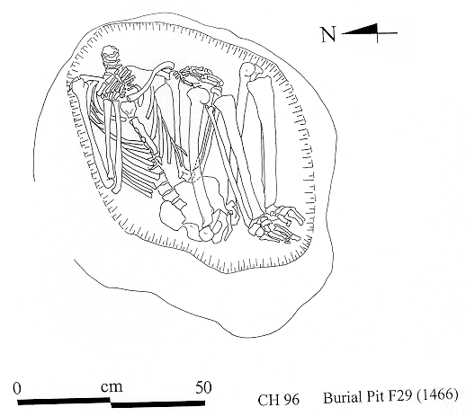
Burial 29
1466 is an articulated skeleton lacking the head and atlas vertebra, adult male.
1467 disarticulated bones of a young adult pushed to one side during burial of 1466, and from same individual, 1470 disarticulated foot bones
1364 hand and foot bones scattered in the upper fill of the grave.
1928 two skulls below 1466, one a juvenile
1949 two skulls and disarticulated bones below 1466
Skeleton 1466
The arms are both tightly flexed on either side of the chest. The legs are also flexed but not acutely, so that the femora diverge from the body and the tibiae also diverge slightly from the femora. There appear to be some phalanges from the feet missing, judging from the drawing (see below).
This specimen provided the best evidence of secondary burial activity, since there is no evidence of disturbance and the skull must have been buried separately, but there is still no indication of any form of defleshing since the skeleton is otherwise complete.
Skeleton 1467
There is some corrosion on many of the epiphyses, e.g. phalanges, radial head. Little corrosion on the much better preserved distal humerus, which at first sight looks different preservation, but in fact it has the same colour differentiation, with manganese restricted to the distal end of the articular surface. There is also some reddish-brown staining on both anterior and posterior surfaces of the distal end of the shaft near the epiphysis that I can not account for. The pelvis has similar preservation again except that being more friable bone anyway it has broken up into irregular friable fragments.
The bones were dispersed on either side of 1466, apparently pushed to one side during this later burial.
CONCLUSION disturbed burial, incomplete
Skeleton 1470
Skeleton 1364
Skeleton 1928
Skeleton 1949
Other material
CONCLUSION
Burial 30
1450, articulated infant buried at the same time as the articulated adult lying on its right side facing the adult.
1426 an articulated hand, femur and vertebrae of an infant
1425 adolescent, the epiphysial condyles haven't been ossified
1464 a single right femur which does not appear to fit any of the above
1494 skull
Skeleton 1424
The body of the articulated adult female (1424) was flexed, the spine very straight, the pelvis rests on the sacrum, lumbar vertebrae face upwards, the thoracic vertebrae are turned so that the right side is presented and the body lies on the left shoulder. For the spine to take on this turn the body must have been placed in the burial position while fresh, whether or not there was subsequent dessication before internment. Grass matting was noted on the outside of the mandible.
All bones are preserved, including both patellae. Some ribs and vertebrae are missing, and some phalanges, but they do not follow any particular pattern. Most of the terminal phalanges are present, and missing are 3 middle phalanges of the hand and one proximal and one middle phalanges of the foot. The sacrum is not present but both sides of the pelvis are preserved.
The ramus of the mandible has several layers of fibrous plant material similar to the phytoliths seen earlier on 1378. The fibre bundles form a network at least three layers thick in one place, running obliquely towards the coronoid notch, but elsewhere on the ramus there are isolated fragments of fibres. Microscopic examination of the rest of the mandible failed to reveal any additional fibre material, and nothing was found on the fragments of face examined.
Fibrous material has also been seen on the ischium, medially on the left humerus, laterally on the olecranon process of the left ulna and medially on the process of the right ulna. Traces occur on the calcaneus and on both femur and tibia shafts but not certain if this is the same thing. None has been seen on the vertebrae, but the outer surfaces of some of the ribs have traces (with black staining on the inner surfaces).
The ribs and thoracic vertebrae have black staining again similar to that seen on1378. The ventral bodies of Thl-7 were all affected and the ribs on both left and right sides articulating with these vertebrae. Cannot tell yet if the staining is on the outer sides of the ribs as well.
Insect channelling is present on the left femur shaft and on some of the ribs, but it is probably recent. There are some recent cut marks from cleaning or excavation, but no old cut marks.
Skeleton 1450
Skeleton 1425
In a separate bag, left and right femur and incomplete tibia and fibula. Preservation is different from 1467, with no trace of manganese staining and the bone is compact and in good condition. There is a skull, complete except for tile base which is shattered. Extensive manganese staining on the outside with traces of phytoliths over the frontal. The interior of the skull has heavy staining on the top of the vault on both frontal and parietals, with the rest of the surface white and no staining at all. Right and left femur with heads separated along epiphyses, extensive black st 9 with white phytolith staining on top of that. Fragmentary pelvis in similar condition, and some blackened ribs with clearly recent insect damage on the surface of two of them.
Skeleton 1426
Skeleton 1464
CONCLUSION
Burial 31
1481 incomplete partially articulated bones from an adult just below floor level and flattened by this proximity;
1482 an adolescent partial skeleton
1483 articulated vertebrae and ribs
1489 skull and vertebrae
1491 six articulated vertebrae in perfect articulation
1498 at the bottom of the pit as presently exposed, an articulated infant
1934 articulated vertebrae and ribs
Skeleton 1481
Included with this is an enormous rib which is not human, and also one bovid incisor. This burial has been disturbed by a large animal hole through the middle of it, and the bones were disturbed by later burials.
Skeleton 1482
Skeleton 1483
In the lower level of the pit (same cut or later one? or even more than one?) are several more partly articulated sets of bones, very incomplete, and one primary burial.
Skeleton 1489
Skeleton 1491
Skeleton 1934
Skeleton 1498
CONCLUSION FOR BURIAL 31
Burial 35
1913 juvenile
1484 juvenile below the first
Skeleton 1913
Skeleton 1484
Burial 36
Skeleton 1495
Burial 38
1922+1939 incompletely articulated skeleton
1923 skull
1924 articulated skeleton
1925 skull and mandible
1928 not known
1926 disarticulated bones
1937 skull
1448 juvenile of about 4 years
1938 partly articulated incomplete skeleton
There is no information on any of these skeletons which are all mixed up together.(see illustration)
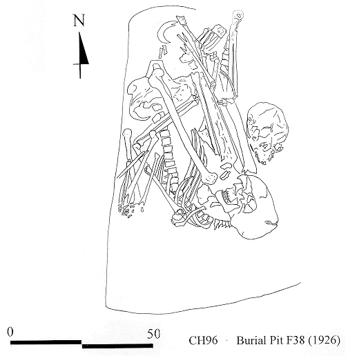
All of the 38 burials, at least 6 skulls and rather more individuals, are disturbed. The surface of this platform had sunk deeply over the burials, being highest to the north edge of the platform by the outside wall. The burial pit went right up to the western wall that divides the room from the end part of the house. It was cut by a large pit that apparently was dug to retrieve something built into the west wall, and parts of two individuals either fell into this pit from the edge of the platform or were actually put there - cannot tell at present. These specimens are labeled 7 on the sketch on page 150. Two amulets were found in front of the neck of the adult.
Skeleton 1448
Burial 40
Skeleton 1912
Skeleton 1950
Burial 41
Skeleton 1940 not known
Burial 42
Burial 44
List of burials
Burial 28
1378 - articulated skeleton of an old adult male
Burial 29
1364 - hand and foot bones scattered on top of pit fill
1466 - articulated skeleton, no head, adult male, final burial
1467 - disarticulated bone of young adult female & 1470 - disarticulated foot bones
1928 - two skulls, juvenile
1949 - two skulls
Burial 30
1424 - articulated skeleton of old adult female
1425 - disarticulated bones: juvenile
1426 - articulated foot, femur, vertebrae of juvenile
1450 - articulated infant, 3-6 months
1464 - femur, right, adult
1494 - skull
Burial 31
1481 - skeleton of adult female
1482 - partial skeleton of adolescent male
1483 - articulated ribs and vertebrae of adult
1489 - 2 partial skeletons
1491 - vertebrae, 5 thoracic, 1 lumbar
1498 - articulated infant intact - latest burial?
1934 - articulated ribs and vertebrae
Burial 35
1913 - articulated child's skeleton, with beads
1484 - articulated child's skeleton
Burial 36
1495 - articulated infant
Burial 38
1922 - incompletely articulated skeleton
1923 - skull
1924 - articulated adult skeleton
1925 - skull and mandible
1926 - disarticulated bones
1937 - skull
193 8 - partly articulated and incomplete skeleton
1448 - Juvenile of 4 years
Burial 40
1912 - articulated infant skeleton
1950 - articulated infant skeleton
Burial 41
1916 - child's skeleton
1940 - not known
Burial 44
193 5 - two juvenile individuals
1474 -juvenile
Unknown burial numbers
Skeleton 1478 - burial ?
Skeleton 1479 - burial ?
Skeleton 1492 - burial ?
Skeleton 1493 - burial ?
Skeleton 1494 - burial ?
Skeleton 1496 - burial ?
| |
© Çatalhöyük Research Project and individual authors, 1996