ÇATALHÖYÜK 1998 ARCHIVE REPORT
    |
Human Remains up to 1998
Theya Molleson, Peter Andrews, Basak Boz,
Jo Sofaer Derevenski, Jessica Pearson
Summary
Isolated fragments of at least three individuals were recovered from Neolithic contexts on the BACH site. In addition the remains of several individuals of probable Roman/Byzantine age were excavated on the BACH site and from a post-Neolithic context on the West mound.
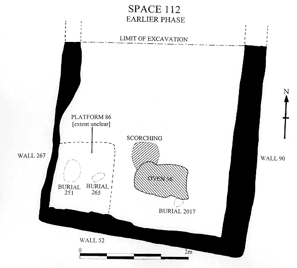
Mellaart Site
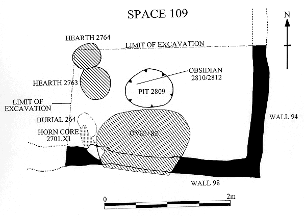
A total of 13 individuals have been excavated from the Mellaart site during the three seasons 1996 (N=0), 1997 (N=10) and 1998 (N=3). The remains of 11 individuals were recovered from Space 112 (Mellaart's Shrine VII.9) including two new-born infants. A third neonate was recovered from Space 109 and a young adult from Space 115.
Inventory of human skeletons from the Mellaart site 1997, 1998:
| Space | Burial | Skeleton | Age | Sex |
| 112 | 83 | 1884 | C, 8y | |
| 112 | 84 | 1885 | C, 7y | |
| 112 | - | 2017 | N | |
| 112 | 84 | 2033 | C, 3y | |
| 112 | 89 | 2056 | MA | M |
| 112 | 89 | 2058 | OA | F |
| 112 | 251 | 2362 | N | |
| 112 | 258 | 2728 | I, 15m | |
| 109 | 264 | 2772 | N | |
| 112 | 265 | 2779 | I | |
| 112 | 274 | 2842 | I, 2y | |
| 112 | 277 | 2886 | MA | M |
| 115 | 285 | 3368 | YA | M |
Descriptions of Human Remains
1884, Space 112, B83
The complete skeleton of an 8 year old child. The child appeared to have a pre-auricular sulcus on each ilium, and cribra femora, both features that could be associated with a habit of sitting cross-legged. The femora show marked torsion of the shaft which is tentatively identified with the habit of sitting with the knees together and the lower legs folded back so that the heels touch the buttocks. (The "mother goddess" position). There is no specific word in Turkish (still less in English) for this position although it is often taken up by the women working in the kitchen. The first metatarsals have extended articulations that are associated with a habit of kneeling with the toes curled under.
1885, space 112, B84
The complete skeleton of a seven year old child. The patella is ossified and fusion of the distal epiphysis of the first metatarsal had started.
Pathology. There is marked cribra in both orbits possibly indicative of long standing anaemia associated with parasite infection (malaria was proposed by Angel 1964). The build up of considerable amounts of calculus on the lingual side of the lower teeth, the incisors especially, and on the buccal side of the upper teeth, regions of salivary ducts, implies a lack of self cleaning or self-cleansing foods and could indicate that the child had been sickly for some time before death.
The child had mild spina bifida occulta with open neural arches of S1, S4 and S5. These neural tube defects have been associated with maternal folic acid deficiency before and during pregnancy. The cribra orbitalia could be linked to this defect.
2017, Space 112, Unit 2013
The partially complete skeleton of a neonatal infant including the skull, part of the spine, tibia and fibula of one leg only.
2033, Space 112, B84
A fairly complete skeleton of a three year old child. Lack of wear on the deciduous molars may indicate that it had not been weaned or if weaned had had a very soft diet.
Pathology. There is moderate cribra of the left orbit (R not observed). The inner table of the right parietal is remodelled near lambda.
Burial 89
2056, Space 112, Burial 89
The underlying body of 2058 was in an extraordinary position, flexed prone, head to south. It is the complete skeleton of a mature-old adult female more lightly built than 2056. The legs were quite tightly flexed at hips and knees but the ankles were extended. The extended feet, the position of the two ilia and the spine indicate that the body had been placed on its front, legs folded under it. The body had soon fallen to its right (west), the head neck and clavicle had displaced to the east and was found in the space between the flexed right femur and tibia of 2056. The right arm had been flexed onto the right shoulder and had essentially remained in place relative to the body. The bones of the right hand were not recovered.
The left arm had become considerably displaced to the east and rotated through about 180o. It was also disarticulated at the elbow and further displaced to the north. The left hand had remained in articulation (all bones were recovered).
A flexed body placed on its knees suggests that 2058 had originally been placed in the grave in a cloth or basket. It is the only case of prone burial so far encountered at the site. Most parts of the skeleton were beneath the larger and more robust bones of 2056, and the displacement of 2058 could have happened at the time of placing 2056 on top incidentally or deliberately to lower the space taken by the remains. The right arm being free at the shoulder, would then have been placed on the edge of the grave cut. There is much heavier staining on 2058 than on 2056, with locally dense manganese staining compared with the moderate overall staining on 2056, and reddish iron staining on 2058 not present on 2056. This could have come about when the grave was reopened to admit 2056, the exposure to air promoting both types of staining on 2058. Some of the black staining on the bones of 2058, notably a band across the left humerus tuberosities may relate to a binding or to clothing, and other places appear to be in concavities where liquids could have pooled. The inside of the skull was white and unstained.
The interval between burial of 2058 and 2056 was not great, though possibly not synchronous if the right arm could be removed so easily; that is unless there was an interval between death and burial of sufficient duration for the limbs to detach readily. This could be a matter of days.
Detailed description of 2056. (Mixing with 2058 possible).
The skeleton of a mature gracile male. The bones of the skull are thick and the sutures are obliterated indicative of an old individual, yet wear on the teeth is slight, especially given the probable age of the individual.
The bodies of all the neck vertebrae are complete; all spinous processes are missing; some facets and transverse processes are fragmented or missing. The joints of the first two cervical vertebrae are notably large. The unciate processes are large on all cervical vertebrae (except C1,C2). The left superior articular facet (SAF) of C3 is distinct and enlarged. The surrounding bone growth is responsive to articulation with the inferior vertebrae. The right SAF is missing. C3-C4, C3 IAF and SAF C4 display severe extensions of the facet margin anteriorly and posteriorly. Extreme asymmetry of osteophyte growth and degeneration to left side. Craters about 1.5mm diameter indicate cartilage resorption. There is eburnation around the anterior margins of the facets. The body displays indications of response to disk pressure. Slight osteophyte growth on inferior margins of the body of C3 in left posterior section. Degenerate change appears to be confined to the mid cervical vertebrae.
The right SAF of the first thoracic vertebra is distinct, the left facets are missing. There is no evidence of disk damage in the lower regions of the spine (ossified ligament on a lumbar joint, possibly L3).
The pelvis has a robust but not protruding inguinal tubercle. The femora are strongly buttressed, have short well marked gluteal tuberosities; Poirier's facets and trochanteric spicules are developed. The distal condyles are medially rotated with respect to the head and neck of the femur.
Squatting facets on the distal right femur and tibia and vastus notching of both patellae, kneeling extensions on right first metatarsal and on both first and second metatarsals of the left foot imply a habit of kneeling with the toes curled under with particular pressure being placed on the left foot.
Pathology. Degenerative changes of the carpo-metacarpo joints of the right second and third metacarpals, with pitting on the right hamate are probably the result of a hand injury.
2058 (B89) Possibly mixed with 2056.
Detailed description
The jaw is small and feminine. There is some evidence of dental reduction with absence of a wisdom tooth.
The lower cervical vertebrae (C4-7) show long standing degenerative changes. The bodies of all vertebrae are complete; all spinous processes are missing, the transverse processes are fragmented. The cervical bodies display indications of severe disk damage or collapse and there is posterior cartilage resorption leading to crater-like depressions on the body and slight eburnation on the superior articular facets (SAFs) and inferior articular facets have fairly distinct margins on both sides. No marked degenerative changes were noted in the thoracic vertebrae, which are generally fragmented however.
The clavicles and scapulae are gracile without degenerative change. The head of the left humerus has slight lipping of the articular margin, the lesser tubercle is pronounced. Distally the olecranon fossa is deep, imperforate and the trochlea ridge marked. The lateral condyle is damaged so that the radius head imprint cannot be observed. The radius and ulna are gracile. Distally the right humerus has a well marked imprint for the radius head and an eburnated groove on the condyle. There are no degenerative changes to the joint. The radius head has a narrower and rolled articular margin where it impacted on the humerus during a compression flexion of the joint. Such a flexion suggests that a heavy load was habitually carried over the shoulder. There is no evident lateral asymmetry of the forearms. The bones of the left hand are small and gracile.
A preauricular sulcus and flared subpubic angle indicate a female. There is a long spicule in the preauricular sulcus - an ossified ligament attachment - and strongly developed inguinal tubercle. These are features associated with multiple births and or a protruding stomach. The public symphysis is old.
The femora are gracile with well developed linea aspera. A distinct Allen's fossa (cribra femoris) on the anterior neck of the left femur and a spicule in the greater trochanter of the right femur suggest habitual rotation of the femora while carrying weight (Angel 1964). The patellae do not have a vastus notch. The tibia-fibula articulation is situated under the proximal articulation which is rolled.
The bones of both feet are present. There is a resorption sinus in the left calcaneus; evidence of the onset of osteoporosis. There is a healed lesion (osteoma) on the shaft of the right third metatarsal, so she may have limped, avoiding putting weight on her foot. The 'kneeling' articulation on the first metatarsal head is only moderate whereas tubercles are well developed on both toes.
In conclusion, 2058 was a woman over fifty, obese, who had been in the habit of carrying heavy loads slung over her shoulder. She had sustained an injury to her right foot at some time and possibly limped slightly. This disuse may have brought on the osteoporosis of the calcaneus. The degenerative changes in the neck vertebrae may be the result of neck strain during carrying; otherwise there are virtually no degenerative changes to any of the joints and she remained mobile to the end of her days.
2362, Space 112, Burial 251.
Neonate or perinatal infant. No cribra orbitalia, although the orbital bones are thick.
2728 space 112, B258
The ribs were deflated in the lower thorax, spaced in the upper thorax, suggesting shallow breathing prior to death.
Pathology. Slight cribra present in both orbits, indicative of anaemia.
2772, Space 109, B264
The well preserved skeleton of a full term neonate.
2779, Space 112, B265
Skeleton 2842, Burial 274, Space 112
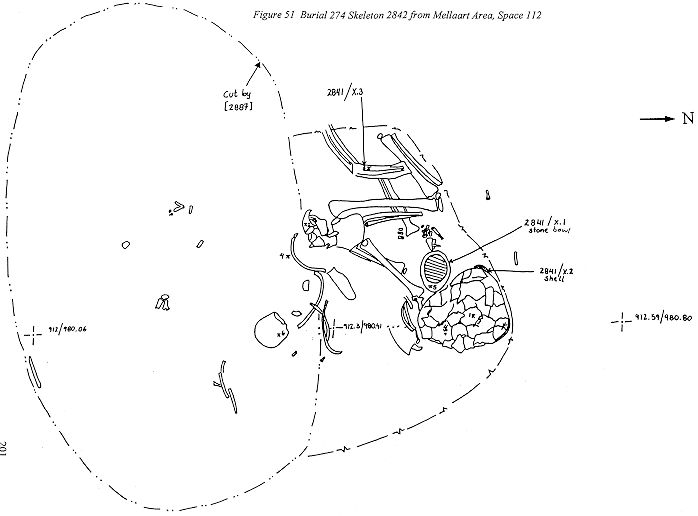
This child was buried with a bowl placed by its head.
Detailed description of the remains
Dental development indicates an age around two to three years (M1 stage 6 Schour and Massler). The dc and dm2 were not fully in occlusion at the time of death. There are contact facets on di1, di2, and dm1.
Occipital lateral wings are not fused and have an unusual suture form which could be familial. Neural arches are fused but not the vertebral bodies. The long bones are in good condition mostly. Epiphyses for the humerus, femur and tibia were recovered. The parts of the proximal humerus epiphyses had not fused at the time of death. The bone maturity is compatible with the dental age.
Skeleton 2886, Burial 277, Space 112
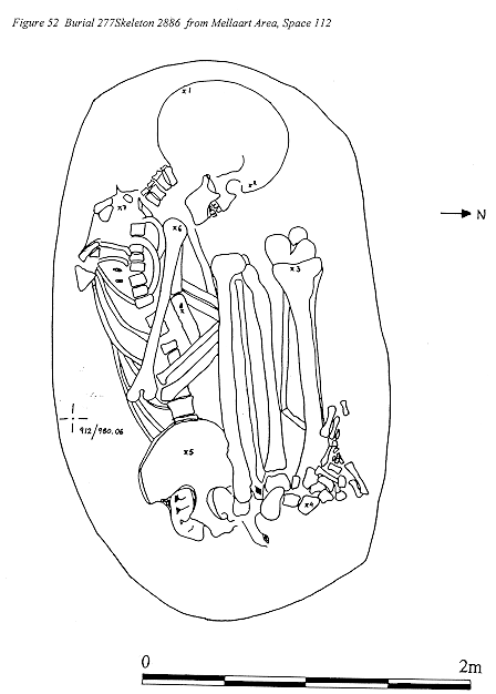
This is the skeleton of a young adult male of very small stature (153cm or 5ft) and marked dental crowding. He appears to have been involved in an accident that resulted in the fracture of a lateral incisor and injury to the upper thorax, involving clavicle, first ribmanubrium, and chest.
The rest of the skeleton shows little evidence of modification apart from mild Scheuerman's asymmetry of the dorsal vertebrae, an asymmetry that can be associated with load bearing in adolescence.
The habitual posture of squatting seems to have been with right leg tightly flexed at the knee and directed to the side of the trunk, the left leg close to the body, tightly flexed at the ankle, resting on the toes. It is not clear whether the torsion of the left femur and rotation of the tibiae is determined by the squatting position or by some other activity.
Detailed description of 2886.
The skull is fragmented and parts are missing so that the face could not be reconstructed. The orbits are notably wider than high with thick well rounded margins (+2), the glabella is inflated, the mastoids large; a characteristic male.
The frontal sinus is small. There is an unusual development of the metopic suture; this extends from bregma for only 40mm towards glabella, which shows only traces of a fused suture. The mastoid is pneumatised. Ossicle at asterion may be familial.
The upper dentition is complete. The third molars are reduced. The right canine is impacted and lies horizontally in the floor of the maxillary sinus. The right I2 had been fractured in life with exposure of the pulp and subsequent apical abscess. There are no dental caries, nor signs of periodontal disease and only slight calculus. Attrition is multifaceted, implying tooth to tooth contact and is possibly task related, since the upper anterior teeth have wear facets the length of the crown palatally.
The lower dentition is complete, with marked anterior crowding and impaction of the lower third molar.
The angle of the jaw is very upright and the gonial angle approaches 90o. The ascending ramus is broad with development of the coronoid. The condyle is very small. This suggests a chewing habit involving the pterygoid muscles which push the jaw forward, rather than the masseter muscles. The inner gonial region is rugose. Overall, attrition is not severe and is equal on both sides, despite the earlier injury and infection.
The hyoid is present and one cricoid. The left first rib has an ossified cartilage to manubrium. Another rib may have a healed crack. Manubriumsternum not fused. Medial articular surface of left clavicle smaller than that of right.
Left innominate is fairly complete and has a narrow sciatic notch (+2) with very narrow sharp edged sulcus of male type. The right public symphysis has young surface.
Vertebrae. C1, C2, C37 have single foramina; strong vertical uncinate processes. Eleven thoracic vertebral bodies were recovered and fragments of arches. Slight Scheuerman's about Th610. Five lumbar vertebrae, with spondylothesis of L5. This dehiscence of the neural arch can have a familial predisposition, set off by load bearing in youth. Sacrum of five vertebrae; S12 fused, hiatus S45.
Both humeri are present. A large supracondylar fossa is present in the right, small in the left. Radial fossa is moderate in right, trace in left. Development of the supinator crest is small in both ulnae. Bones of hands show that there was no marked handedness or robusticity.
Right and left femora complete. Torsion of left is greater than that of right; no Poirier's or Allen's facet; no trochanteric spicule; very platymeric; strong gastrocnemius attachment (stronger in left). Patellae both have vastus notches.
Right and left tibiae. Right has rounded lateral platform of squatting but distally lateral squatting facet much larger on left. There is marked torsion between proximal and distal tibiafibula articulation, so he was pigeon toed or turned his knees out. Proximal tibiafibula articulation under articular platform indicating weight bearing fibula. Not platycnemic.
Foot bones include first metatarsal with kneeling articulation and kneeling tubercle on lateral side.
Skeleton 3368, Burial 285, cut 3369, Space 115 Midden deposit. (Not seen by PA)
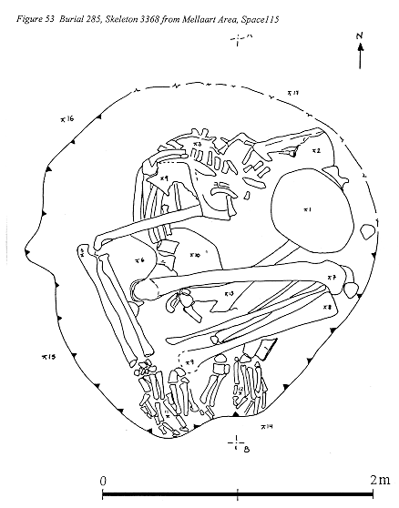
There was no disarticulation of any joint. The upper (right) side had been displaced caudally relative to the left, so that the two scapulae were situated right below left and right ilium below left. The right arm was bent at the elbow about 90o, yet the forearm lay below the ilium and hand over the bones of the right foot, an impossible position in life. The elbow joint must have flexed, subsequent to burial, presumably at the time of the caudal movement of the right scapula. The head had tipped down and the patellae were both in articulation at the top edge of the pit.
The lie of the surface, as revealed by the excavators, dropped from head to hip, so that the body can be interpreted as having collapsed downhill. The skull was crushed but contained soil. Ilium, radius and ulna had 'old' pressure cracks; the tibia had longitudinal breaks; the femur had breaks at each end but not the shaft, suggesting that compacting pressure was not great. The pit appeared to be a flat scrape in the midden floor. The bones are particularly darkly stained.
On the sacrum and between the ilia were patches of pale orange substance containing hackberry seeds. If this material is coprolite it implies a relatively sudden death; no long illness with fever during which the individual would not have eaten.
This is the complete, articulated skeleton of an adult male in his early 20s. Although recovery was good many of the bones are very friable and severely fragmented, especially the vertebrae. It proved impossible to separate bones at some joints. This may result from compaction in the soil but may also be related to the severely porotic condition of many of the bones of the trunk and shoulders. 3368 is the only case of chronic, systemic bone pathology, that has been recovered either from the current or from the original excavations.
Detailed description of the remains
The skull bones are fragmented but apparently normal apart from a small circular lesion on the right parietal near the sagittal suture. The bone around this is porotic. The mastoids are asymmetric. Left maxilla (right not recovered) had lost all the anterior teeth, I1P2 long before death the alveolar bone is totally resorbed and the palate is flat. In the mandible the anterior teeth, LI1RI2 had also been lost antemortem. A healed abscess under I2 suggests that this may result from an injury. Dentally the teeth that survive are healthy though well worn for the age at death.
The vertebrae and ribs were largely too fragmented for recovery; some bear evidence of bone disease.
The scapulae are grossly pathological with swollen coracoid, acromion and spine especially on the left. The articulation of the glenoid is not affected. Clavicle sharply angled (this may be familial cf North 1995) and pathological.
The right humerus has a larger midshaft diameter and Dshaped cross section, whereas that of the left is oblong. The right has a large supracondylar foramen; the left is damaged but a small foramen was present. The radial imprint is not strong.
The right ulna is swollen in the distal third and has a strong but short supinator ridge. the line of the distal epiphysis is still patent. The left ulna presents a severe periostitis of the shaft especially of the proximal and distal shaft where the olecranon is broken off. The inner bone is coarsely spongy throughout. Around this break, which is postmortem, there is a thick surface periostitis. Along the distal shaft the periostitis presents as a thick crust tending to flake off. The carpal bones of both hands look normal.
Metacarpals III, IV and V of the right hand show evidence of periostitis. MC V has a swollen shaft with porotic surface. The articulation is not affected. The proximal phalanges also show some periostitic changes; the Vth is swollen. Of the medial phalanges phalanx III has osteitis distally, possibly on the site of injury; Ph IV and V are porotic. The terminal phalanges are also affected.
In the left hand MC II is swollen and deformed distally; the articulations are not affected. This bone is much shorter than that of the right (58.2, 63.7). MC III, IV, V are also porotic. Of the proximal phalanges I and III have periostitis. The right ilium has porotic changes around the crest, and the pubic symphysis is porotic.
The right femur shaft and head appear normal, as do the patella and tibia but the fibula ends are pathological. The tarsal bones and metatarsals are very friable. The phalanges are porotic.
The proximal phalanx of the big toe has a kneeling angle which is less than that of the left big toe, suggesting a squatting habit that was asymmetrical.
Pathology. The youth had suffered from a chronic systemic and probably painful illness for some time. The disease had affected the bones of the shoulder, spine, ribs and hips in particular, but also individual bones of the forearm (ulnae) and hands. The bone is highly abnormal, swollen and spongy. The ribs appear to have fractured easily; some fractures have healed with weak callus formation, other fractures have failed to unite. The disease has also located around a minor injury to the terminal joint of the middle finger. In general though the joint surfaces are not involved.
A gross superficial periostitis has developed along the shaft, both proximal and distal half, of the left ulna. This too could be the site of an injury or a response to pulmonary problems (idiopathic pulmonary hyperostosis).
Apart from a small lesion on the top of the cranium, the head and face were apparently not involved in the disease process, although the antimortem loss of so many teeth at such a young age might be due to the disease.
Figure 51: Burial 274 Skeleton 2842 from Mellaart Area, Space 112
Figure 52: Burial 277Skeleton 2886 from Mellaart Area, Space 112
Figure 53: Burial 285, Skeleton 3368 from Mellaart Area, Space 115
BACH Area
In the 1998 season isolated human bones were excavated from a two metre square area, in Neolithic levels. The remains include: calotte; several vertebrae; two innominate bones; humerus and fibula . These are presumed to belong to a single adult female. Also the right parietal of an infant which, based on size, would have been about six months at the time of death. In addition there were also bones from a juvenile aged about 6 years.
Inventory of human skeletal material from BACH
| Number | Feature | Age | Sex | Artifacts |
| Byzantine (5-6th century) | ||||
| 2210 | Adult | burnt | ||
| 2212+2231 | F151 | Adult | F | glass bottle |
| 2219 | F150 | Adult | M | |
| 2226+ | F152 | Juv, 4y | Blue bottle | |
| 2232 | Cu beads, needle | |||
| 2235+2245 | F153 (Gr5) | Adol, 16y | M? | |
| 2244 | F154 | Adult | M | |
| 2265 | Adult | M | ||
| Neolithic | ||||
| 2229 | Adult | |||
| 2255 | Adult | |||
| 2255 | F149 | Juv, 6y | ||
| 2263 | Adult | |||
| 2270, 2281 | Adult | F | ||
| 2281 | Infant | |||
| 2293 | Adult | |||
Detailed Description of Byzantine Remains.
2210
Feature 150 Associated units 2205, 2206, 2219
Feature 151 Associated units 2211, 2212, 2231
Feature 152 Associated units 2225, 2226, 2232
Feature 153 Associated units 2234, 2235, 2245
Late adolescent-young adult male skeleton. Remains are very fragmented. The feet from this skeleton excavated in 1997 were subsequently recovered during the 1998 season. The calcaneus epiphysis had already fused, though some epiphyses in the remainder of the skeleton were only in the process of fusing. Kneeling articulations were observed, though the lack of squatting facets on either the tibiae or tali argues against squatting as a habitual posture. Flattening of MTII head and kneeling articulation could indicate a kneeling habit with heels off the ground.
Feature 154 Associated units 2236, 2237, 2244
2265
Details. Skull with antemortem loss of molars, occlusal calculus on lower M3. Hyoid and ossified thyroid (indicating buried articulated). Right ribs with black staining on inside and on upper side of rib 1. Manubrium fragment, sternum with ossified xiphisternum. Scapulae, sacrum, ilia, vertebrae including C16, thoracic and lumbar, without signs of loadbearing. Arm, L,R femur heads, tibia head, hand and foot bones very fragmented.
Neolithic Skeletal Remains
2229, F285:
2251
2255 F963:
F149: Juvenile proximal third left femur, with unfused epiphysis, cribra femoris. G: tooth germ upper left canine crown complete (= 6 years). NB has apparently worn tip but certainly not erupted; hypoplastic lines at base of crown.
2257
2263
2270
2281
2281
Also scapula which has different preservation and looks like Roman.
2281
2293
West Mound
2901
2909 + 2916
2913
2918
2920
2922I
2922II
2922III
Conclusion
The burial assemblage which was distributed throughout the excavated units appears to consist of the remains of a minimum of two adult individuals one of which was probably male. The measurements which could be taken from the remains show close resemblance to other Byzantines studied from Turkey*, however, since there are no Chalcolithic remains for comparison at present, the assignment of a Byzantine date according to measurements taken from the bones should not be considered conclusive. The burials were clearly disturbed. This can be supported from the lack of patellae and femora and much of the axial skeleton. The skulls could, potentially, be Chalcolithic (according to the archaeology). Measurements could not be taken of the surviving crania, it would be difficult to assess a difference in population type between the skeletal remains recovered from such a small population sample. The alternative would be to use the radiocarbon method of dating.
*Domuztepe unpublished skeletal analysis, 1998.
| |
© Çatalhöyük Research Project and individual authors, 1998